Iris and Fundus Coloboma: Causes, Symptoms & Modern Eye Care Management
anushka
18 November 2025
Eye Health
Iris and fundus coloboma are uncommon yet important developmental eye conditions that every parent, patient, and eye-care professional should understand. In many cases, a coloboma is identified during childhood, but some patients remain undiagnosed until adulthood—often presenting with decreased vision, glare, or cosmetic concerns.
This blog explains what coloboma is, why it happens, how it affects vision, and what modern treatments are available. It is written in easy clinical language suitable for patient education and hospital websites.
What is a Coloboma?
A coloboma is a structural defect in the eye present since birth. It occurs when a specific tissue gap—called the embryonic fissure—fails to close properly during early fetal development (around 5–7 weeks of gestation).
Depending on where this gap remains open, different parts of the eye may be affected:
Iris coloboma
Lens coloboma
Choroidal coloboma
Optic disc coloboma
Retinal (fundus) coloboma
Some patients may have one type, while others may have a combination.
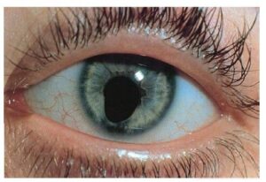
Iris Coloboma
What is Iris Coloboma?
It is a keyhole-shaped or notch-shaped defect in the iris. The pupil may look elongated or “pear-shaped” downward.
Symptoms of Iris Coloboma
Light sensitivity (photophobia)
Glare, especially in sunlight
Cosmetic concerns
Sometimes mild vision reduction if the defect is large
How It Affects Vision
The iris controls the amount of light entering the eye. A coloboma makes the pupil larger or irregular, allowing excessive light inside — causing glare and reduced contrast sensitivity.
Treatment Options
Iris coloboma cannot be reversed, but symptoms can be managed:
1. Cosmetic Contact Lenses
Special iris-print lenses can mask the appearance and reduce glare.
2. Sunglasses / UV-Protection Glasses
Help manage glare and photophobia.
3. Artificial Iris Implant (in selected cases)
A surgical option for severe defects or trauma-related enlarged pupils.
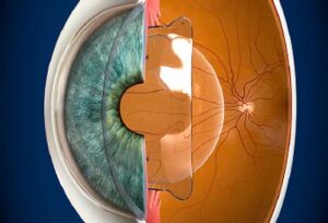
Fundus (Choroidal/Optic Disc) Coloboma
What is Fundus Coloboma?
Fundus coloboma occurs when the retina, choroid, or optic nerve fails to form completely. It appears as a sharply demarcated white or pale area during a dilated fundus examination.
Types:
Choroidal coloboma (most common)
Optic disc coloboma
Combined coloboma
Symptoms of Fundus Coloboma
Symptoms depend on the size and location of the defect:
Blurred or reduced central vision
Missing visual field areas
High myopia or astigmatism
Increased risk of retinal detachment
Strabismus (squint) in children
Nystagmus in severe cases
Why Fundus Coloboma is Important?
These eyes are more prone to complications, especially:
1. Retinal detachment
Because the thinned-out retina near the coloboma is fragile.
2. Choroidal neovascularization (rare)
Abnormal blood vessel formation.
3. Amblyopia in children
If not treated early with glasses/patching.
Causes of Coloboma
Coloboma is congenital (present at birth). Main causes include:
Genetic mutations
Chromosomal abnormalities (like CHARGE syndrome)
Family history of coloboma
Developmental interruption during pregnancy
Most cases are sporadic and not caused by anything the parents did.
Diagnosis
How is Coloboma Diagnosed?
✔ For Iris Coloboma
Torchlight exam
Slit-lamp biomicroscopy
Photographic documentation
✔ For Fundus Coloboma
Dilated fundus examination
OCT (to study involvement of macula)
Fundus photography
Visual field testing
Ultrasound in unclear cases
Treatment & Management
1. Glasses / Spectacles
Correct refractive errors like myopia, hyperopia, or astigmatism.
2. Amblyopia Therapy (in children)
Glasses
Patching of the better eye
Vision therapy (depending on the case)
3. Monitoring for Retinal Detachment
Regular retinal check-ups are essential.
4. Low Vision Aids
For patients with significant visual field loss or macular involvement.
5. Surgery
While the coloboma itself cannot be completely “fixed,” certain surgeries help with its effects:
Artificial iris implants
Cataract surgery modifications for lens coloboma
Repair of retinal detachment if it occurs
Prognosis: How is the Vision in Coloboma Patients?
Vision depends on:
Size of the coloboma
Whether the macula is involved
Presence of complications (like detachment)
Early intervention for amblyopia in children
Many patients maintain useful day-to-day vision with proper management and protective care.
How Can Patients Protect Their Eyes?
Use UV-protected sunglasses
Regular retina check-up every 6–12 months
Immediate consultation if symptoms like flashes, floaters, or sudden vision drop appear
Protective eyewear for children
Full-time glasses use where indicated
Conclusion
Iris and fundus coloboma are congenital eye conditions that require lifelong monitoring, but with advances in diagnostics, vision therapy, and protective strategies, patients can maintain stable vision and enjoy good quality of life.