Anushka Super Speciality Eye Hospital

Diseases of the Pupil: Causes, Systemic Associations, and Clinical Insights

Eye Health

Introduction

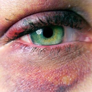
The pupil, the dark circular opening in the center of the iris, plays a vital role in regulating the amount of light that enters the eye. Its size and reaction to light provide critical information not only about ocular health but also about neurological and systemic well-being. Abnormalities in the pupil—whether in size, shape, symmetry, or reactivity—often indicate underlying disease processes.

This blog explores the diseases of the pupil, their causes, clinical significance, systemic associations, and management strategies, offering a detailed overview for students, clinicians, and health-conscious readers.

Anatomy and Physiology of the Pupil

The pupil functions like a camera aperture:

  • Dilates in dim light to allow more light into the eye.
  • Constricts in bright light to protect the retina.

This reflex is controlled by two muscles within the iris:

  • Sphincter pupillae (parasympathetic control) → causes constriction.
  • Dilator pupillae (sympathetic control) → causes dilation.

Therefore, pupil abnormalities may indicate dysfunction of the ocular structures, autonomic nervous system, or systemic disease.

Categories of Pupil Abnormalities

Diseases of the pupil can be grouped into abnormalities of:

  1. Size – small (miosis) or large (mydriasis).
  2. Shape – irregular pupils.
  3. Equality – anisocoria (unequal pupil sizes).

Reactivity – sluggish, absent, or paradoxical light response.

Major Pupil Disorders and Their Causes

  • 1. Anisocoria (Unequal Pupils)

    • Defined as a difference in pupil size >0.4 mm.
    • Can be physiological (benign, present in 20% of population) or pathological.

    Causes:

    • Horner’s syndrome (sympathetic damage → small pupil, ptosis, anhidrosis).
    • Adie’s tonic pupil (parasympathetic denervation → large, sluggish pupil).
    • Cranial nerve III palsy (compressive lesion → dilated pupil with ptosis and ophthalmoplegia).
    • Pharmacological effects (eye drops, drugs).

    2. Miosis (Small Pupil)

    Defined as persistent constriction of the pupil (<2 mm).

    Causes:

    • Physiological in infants and elderly.
    • Pharmacological: pilocarpine, opioids.
    • Neurological: Horner’s syndrome, pontine hemorrhage.
    • Inflammatory: uveitis with posterior synechiae.

    Systemic association: Opioid overdose is a key systemic cause, where “pinpoint pupils” are a diagnostic hallmark.

    3. Mydriasis (Dilated Pupil)

    Abnormally enlarged pupils (>6 mm).

    Causes:

    • Traumatic mydriasis (iris sphincter tear).
    • Pharmacological: atropine, tropicamide, sympathomimetics.
    • Neurological: third nerve palsy, brain herniation (fixed dilated pupils = life-threatening).

    Systemic association: Raised intracranial pressure (ICP) or uncal herniation often presents with a dilated, non-reactive pupil—an emergency sign.


    4. Irregular Pupil (Dyscoria)

    The pupil loses its round shape.

    Causes:

    • Anterior uveitis → posterior synechiae pulling the pupil.
    • Trauma or surgery (iris tears).
    • Congenital coloboma (keyhole-shaped pupil).

    5. Light-Near Dissociation

    A condition where the pupil does not respond to light but constricts on near effort.

    Causes:

    • Argyll Robertson pupil → seen in neurosyphilis, famously described as “accommodates but does not react.”
    • Adie’s tonic pupil → light reaction absent, near response slow and tonic.
    • Dorsal midbrain lesions (Parinaud’s syndrome).

    6. Relative Afferent Pupillary Defect (RAPD / Marcus Gunn Pupil)

    • Occurs when one optic nerve transmits less light input than the other.
    • Detected with the swinging flashlight test.

    Causes:

    • Optic neuritis (common in multiple sclerosis).
    • Severe retinal disease (retinal artery occlusion, detachment).

Systemic Diseases Associated with Pupil Abnormalities

The pupil often reflects systemic illness. Some important associations include:

Neurological Disorders

  • Horner’s Syndrome: Associated with brainstem stroke, carotid artery dissection, apical lung tumors (Pancoast).
  • Third Nerve Palsy: Caused by aneurysms, trauma, or diabetes.
  • Multiple Sclerosis: May present with RAPD due to optic neuritis.

Infectious Diseases

  • Neurosyphilis: Causes Argyll Robertson pupil.
  • Herpes Zoster Ophthalmicus: Can cause irregular pupil due to iris atrophy.

Metabolic and Drug-Related Conditions

  • Diabetes mellitus: Can cause autonomic neuropathy affecting pupil response.
  • Drug intoxication: Opioids (miosis), cocaine/amphetamines (mydriasis).

Critical Illness and Emergency Conditions

  • Head trauma / brain herniation: Fixed, dilated pupils are ominous.

Pontine hemorrhage: Causes pinpoint pupils.

Diagnostic Approach to Pupil Disorders

  1. History: Onset, trauma, drug use, systemic disease.
  2. Examination: 
    • Size, symmetry, and reactivity to light/near.
    • Presence of ptosis, anhidrosis (Horner’s).
    • Ocular motility (nerve palsy).
  3. Special Tests: 
    • Swinging flashlight test (RAPD).
    • Pharmacological testing (e.g., apraclonidine for Horner’s).
    • Neuroimaging for suspected neurological causes.

Management of Pupil Disorders

Treatment depends on the underlying cause:

  • Pharmacological causes: Discontinue offending drugs.
  • Inflammatory causes (e.g., uveitis): Steroid drops, cycloplegics.
  • Neurological causes: Manage stroke, aneurysm, tumor, or infection appropriately.
  • Horner’s syndrome: Treat underlying pathology (e.g., carotid dissection).
  • Third nerve palsy with pupil involvement: Urgent neuroimaging for aneurysm.
  • Traumatic mydriasis: May require surgical repair if persistent.

Prognosis

  • Benign conditions like physiological anisocoria are harmless.
  • Systemic or neurological causes may be life-threatening if not recognized early.
  • Timely management often improves visual and systemic outcomes.

Prevention and Public Awareness

  • Regular eye check-ups can detect subtle pupil abnormalities.
  • Patients with diabetes, hypertension, or neurological symptoms should undergo periodic eye exams.
  • Awareness of warning signs like sudden anisocoria, fixed dilated pupil, or painful third nerve palsy is crucial for early medical intervention.

Frequently Asked Questions (FAQs)

1. What does it mean if one pupil is bigger than the other?
Mild differences are normal, but sudden anisocoria may indicate neurological emergencies like Horner’s syndrome or third nerve palsy.

2. Can stress or emotions affect pupil size?
Yes. Pupils dilate during stress or emotional arousal due to sympathetic activation.

3. Which drugs affect pupil size?
Opioids cause pinpoint pupils, while cocaine, amphetamines, and anticholinergics cause dilated pupils.

4. Are pupil diseases always serious?
Not always. Some are benign, but others may indicate stroke, aneurysm, or systemic illness.

5. Can pupil abnormalities cause blindness?
By themselves, no—but they often signal diseases (like optic neuritis or retinal detachment) that can lead to vision loss.

Conclusion

The pupil is more than just a gateway for light—it is a window into systemic health. Disorders of the pupil reflect a wide spectrum of conditions, ranging from harmless physiological variations to life-threatening emergencies.

A careful assessment of pupil size, shape, and reactivity provides valuable clues in diagnosing neurological, infectious, and systemic diseases. For clinicians, pupils remain an essential diagnostic tool. For patients, awareness of abnormal pupil signs and seeking timely care can be sight-saving and even life-saving.

Tags :
Eye Health
Share This :